Medically reviewed for current US clinical guidance · Last reviewed: July 16, 2026
Breast cancer in the United States arises from a complex interaction of genetics, hormones, lifestyle, and environmental factors. While most cases happen with no clear single cause, several factors are well-established as raising risk. Diagnosis usually starts with screening mammography or symptoms, then progresses through diagnostic imaging, biopsy, and staging. This article walks through what is known about causes, risk factors, and the diagnostic process.
If you are new to breast cancer, the cluster overview article is a useful starting place.
What Causes Breast Cancer?
Breast cancer develops when cells in the breast acquire DNA mutations that allow them to grow uncontrollably. The exact triggers usually cannot be identified for an individual patient. Contributing mechanisms include:
Hormonal influences. Lifetime exposure to estrogen affects breast cell behavior. Factors like early menstruation, late menopause, and certain hormone therapies extend hormone exposure and raise risk.
Inherited genetic mutations. Mutations in BRCA1, BRCA2, and other genes (PALB2, TP53, CHEK2, ATM, PTEN, and others) significantly raise lifetime risk. These account for about 5-10 percent of breast cancer cases.
Acquired DNA damage. Random mutations accumulate over time. Some are influenced by environmental factors like radiation exposure or certain chemicals.
Cell growth signaling. Errors in cell growth controls (HER2 overexpression, hormone receptor signaling) drive specific cancer subtypes.
Inflammation and obesity. Chronic inflammation and obesity-related hormone effects can contribute, especially post-menopause.
For most patients, breast cancer arises from a combination of multiple factors over time. Most patients did nothing “wrong” to cause their cancer.
Major Risk Factors for Breast Cancer
Several factors significantly raise breast cancer risk in US adults.
Non-modifiable factors:
- Female sex (women have ~100 times the risk of men)
- Age (risk rises steadily after 40; most diagnoses in women over 50)
- Family history of breast or ovarian cancer (especially first-degree relatives)
- Inherited genetic mutations (BRCA1, BRCA2, PALB2, TP53, others)
- Personal history of breast cancer or atypical breast tissue
- Dense breast tissue
- Early menstruation (before 12) or late menopause (after 55)
- No pregnancies or first pregnancy after 30
- Prior chest radiation (e.g., for childhood lymphoma)
- Race/ethnicity differences (different patterns of incidence and aggressiveness across groups)
Modifiable factors:
- Postmenopausal obesity
- Sedentary lifestyle
- Alcohol consumption (risk rises with intake)
- Hormone replacement therapy (combined estrogen-progestin)
- Smoking
- Not breastfeeding (breastfeeding modestly reduces risk)
The combined effect varies. Many women with several risk factors never develop breast cancer; many women with no obvious risk factors do.
Common Symptoms That Suggest Breast Cancer
Symptoms warranting prompt evaluation:
- New lump or thickening in breast or armpit
- Change in size or shape of one breast
- Skin dimpling, puckering, or texture change
- Nipple inversion that’s new
- Nipple discharge (especially bloody or one-sided)
- Persistent redness, scaliness, or “orange peel” skin
- Persistent breast pain in one location
Note: most breast lumps are benign. But any new or persistent change warrants evaluation.
US Breast Cancer Screening Guidelines
The US Preventive Services Task Force (USPSTF), American Cancer Society (ACS), and other US organizations have screening recommendations. As of 2024:
- USPSTF: Recommends biennial screening mammography for women ages 40 to 74 at average risk.
- ACS: Recommends annual mammography starting at age 45 (with option from 40) and continuing at least to 54, then biennial thereafter or annual if preferred.
- High-risk women: May benefit from earlier and/or additional screening (MRI), often starting in their 20s or 30s.
Specific recommendations may vary by your medical history; discuss with your primary care doctor or breast specialist.
How US Clinicians Evaluate Suspected Breast Cancer
The standard US workup for suspected breast cancer typically progresses through screening, diagnostic imaging, biopsy, and staging.
Clinical history and physical examination
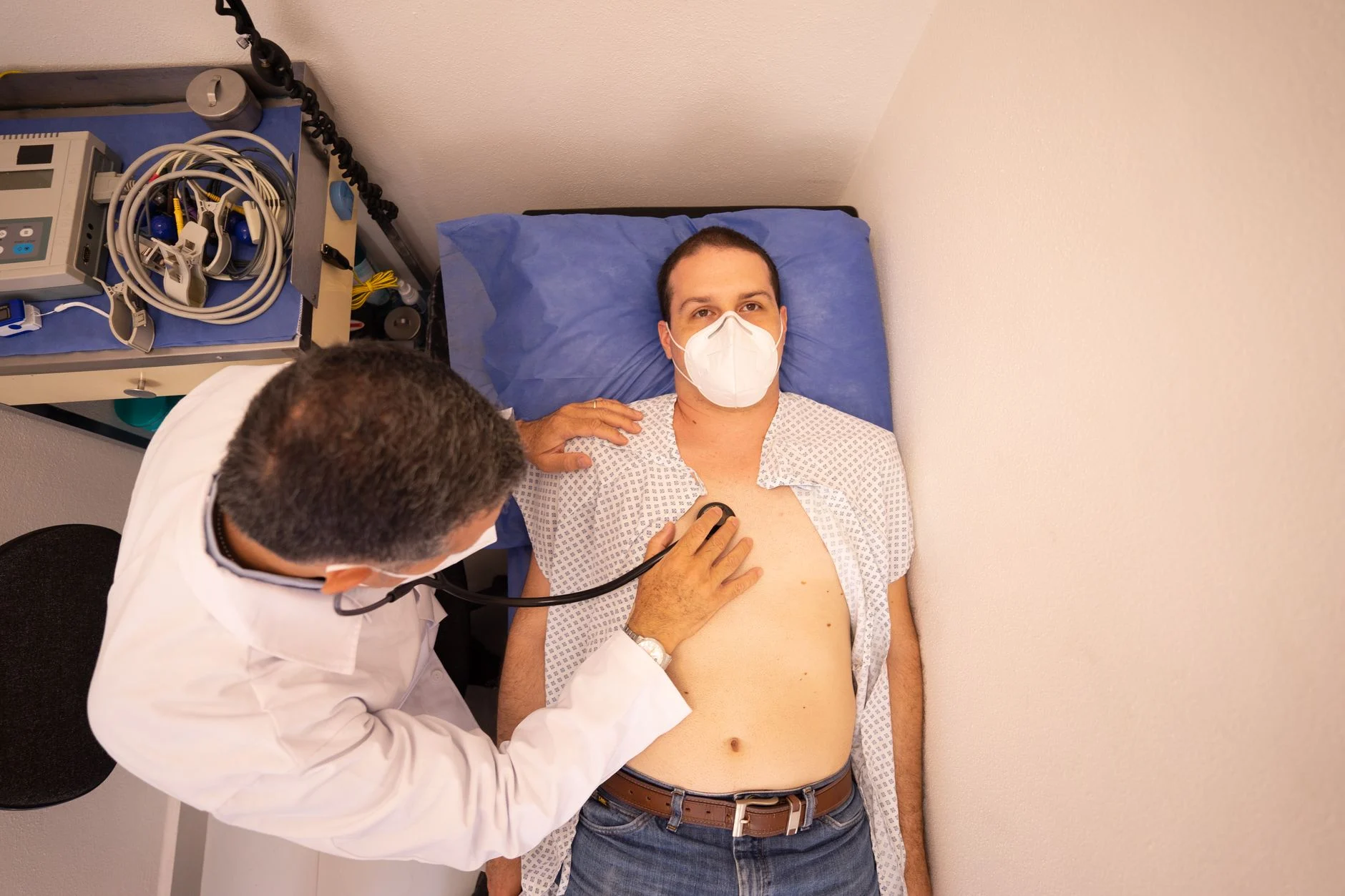
Questions about symptoms, timing, family history, hormonal history, prior biopsies, medications, and breast/body changes. Physical exam includes inspection and palpation of both breasts and lymph nodes (armpit and collarbone).
Diagnostic imaging
Diagnostic mammography. More detailed than screening mammography; includes additional views and magnification.
Breast ultrasound. Distinguishes solid masses from cysts; especially useful in dense breasts or for further evaluation of mammographic abnormalities.
Breast MRI. Highly sensitive; used in selected cases (high-risk women, problem-solving, evaluation of newly diagnosed cancer extent, breast implant evaluation).
Other imaging. Sometimes contrast-enhanced mammography or specialized techniques.
Biopsy
The only way to confirm cancer is by biopsy — removing tissue for microscopic examination.
- Core needle biopsy — the most common technique; tissue cores taken under image guidance
- Vacuum-assisted biopsy — for smaller or calcified lesions
- Surgical (excisional) biopsy — less common but used when needle biopsy is not feasible
- Fine needle aspiration (FNA) — sometimes for lymph nodes or cysts
The pathologist examines the tissue and determines:
- Type of cancer (ductal, lobular, etc.)
- Grade (how aggressive the cells look)
- Hormone receptor status (estrogen ER, progesterone PR)
- HER2 status
- Other markers (Ki-67, others)
Staging
After diagnosis, staging assesses the extent of disease.
- Clinical staging: Physical exam plus imaging
- Pathological staging: Based on surgical pathology of tumor and lymph nodes
- TNM system: Size of Tumor, Lymph Node involvement, Metastasis
Staging may include:
- CT scan of chest, abdomen, pelvis
- Bone scan
- PET-CT
- Brain MRI (if symptoms)
- Lymph node biopsy (sentinel or axillary)
- Blood tests (CBC, liver function, others)
Genetic counseling and testing
For patients with family history suggestive of inherited cancer syndromes, personal history of certain cancers, or younger age at diagnosis:
- BRCA1 / BRCA2 testing
- Panel testing for multiple genes (PALB2, TP53, ATM, CHEK2, others)
- Counseling about implications for treatment, future risk, and family members
Differential Considerations
Not every breast change is cancer. The diagnostic workup may identify or rule out:
- Fibrocystic changes
- Fibroadenoma (benign tumor)
- Cysts
- Mastitis (breast inflammation/infection)
- Lipoma
- Phyllodes tumor (uncommon)
- Other rare breast tumors
The pathology results guide further care.
When to See a Doctor
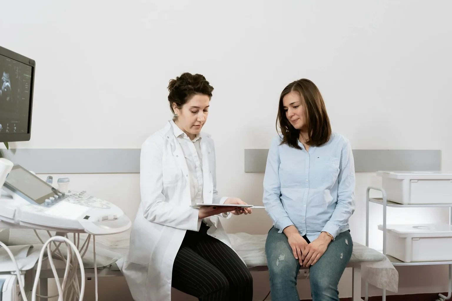
Schedule an evaluation with your primary care doctor or breast specialist if you:
- Find a new breast lump or thickening
- Notice a change in breast size, shape, or skin
- Have new nipple inversion or discharge
- Have persistent breast or nipple pain
- Are at high risk and not currently being screened
- Are not sure when to start screening
Seek prompt evaluation if changes persist more than 2-4 weeks.
How the Diagnosis Is Made
The diagnosis of breast cancer typically follows this process:
- Screening mammography identifies abnormality OR symptoms prompt evaluation
- Diagnostic imaging (mammography, ultrasound, sometimes MRI) characterizes the abnormality
- Biopsy confirms or rules out cancer
- Pathology determines type, grade, ER/PR/HER2 status
- Staging tests assess extent of disease
- Multidisciplinary team review (medical oncology, surgical oncology, radiation oncology) develops treatment plan
- Genetic counseling when indicated
What Happens After Diagnosis
Once breast cancer is diagnosed, the multidisciplinary team develops an individualized treatment plan based on:
- Type and grade
- Stage
- Receptor status (ER, PR, HER2)
- Patient health and preferences
- Genetic findings
- Reconstructive preferences (if mastectomy is needed)
Treatment options are covered in our treatment article and our mastectomy cluster for surgical pathway.
Continue Reading the Breast Cancer Cluster
- Breast Cancer: Overview, Types, and Symptoms
- Breast Cancer: Treatment, Prevention, and Management
- Breast Cancer: FAQs, Statistics, and Patient Stories
- Mastectomy: Causes, Diagnosis, and When to Consider Surgery
Sources
- American Cancer Society (ACS). Breast cancer screening and diagnosis. https://www.cancer.org/cancer/types/breast-cancer.html
- National Cancer Institute (NCI). Breast cancer diagnosis. https://www.cancer.gov/types/breast
- U.S. Preventive Services Task Force (USPSTF). Breast cancer screening recommendations. https://www.uspreventiveservicestaskforce.org/
- Centers for Disease Control and Prevention (CDC). Breast cancer. https://www.cdc.gov/cancer/breast/
- National Comprehensive Cancer Network (NCCN). Breast cancer guidelines. https://www.nccn.org/
- Susan G. Komen Foundation. Breast cancer information. https://www.komen.org/
- Mayo Clinic. Breast cancer diagnosis. https://www.mayoclinic.org/diseases-conditions/breast-cancer/diagnosis-treatment/drc-20352475
- Cleveland Clinic. Breast cancer. https://my.clevelandclinic.org/health/diseases/3986-breast-cancer
Medical Disclaimer
The information in this article is for general education and is not a substitute for professional medical advice, diagnosis, or treatment. Always consult a qualified medical professional with questions about breast cancer, screening, or testing. Any new breast lump, persistent change, or nipple discharge warrants prompt evaluation.