Gallstones are common in the United States, but the underlying causes are surprisingly varied: genetics, weight, diet, hormonal status, certain medications, and underlying medical conditions all contribute. When symptoms develop, a sequence of clinical evaluation and imaging tests is used to confirm the diagnosis and plan treatment. This article walks through what causes gallstones, who is at higher risk, how US clinicians evaluate the patient, and how the diagnosis is made.
If you are new to gallbladder disease, the cluster overview article is a useful starting place.
What Causes Gallstones?
Gallstones form when the chemical balance of bile shifts in a way that allows hardened deposits to develop. The two main mechanisms in US patients are:
Excess cholesterol in bile. When the liver secretes more cholesterol than bile can dissolve, cholesterol crystallizes and forms stones over time. This is the most common cause and produces the typical yellow-green cholesterol gallstones found in approximately 80 percent of US cases.
Excess bilirubin in bile. When the body produces too much bilirubin (a breakdown product of red blood cells), small dark pigment stones can form. Common in patients with chronic blood disorders, cirrhosis, or biliary infections.
Other contributing mechanisms include:
- Gallbladder dysfunction. When the gallbladder does not empty completely or often enough, bile becomes concentrated and prone to stone formation.
- Reduced bile salts. Lower levels of bile salts (the natural emulsifiers in bile) leave cholesterol less soluble.
- Mucus and inflammation. Chronic inflammation can change the gallbladder’s chemistry.
Major Risk Factors for Gallstones
Several factors significantly raise the likelihood of developing gallstones in US adults.
Demographic factors:
- Sex: Women are about twice as likely to develop gallstones as men, largely due to estrogen effects on bile cholesterol.
- Age: Risk rises sharply after age 40.
- Ethnicity: Higher rates in Hispanic and Native American populations.
- Family history: Gallstones tend to run in families.
Body weight and metabolism:
- Obesity (high BMI): Strongly associated with cholesterol gallstones.
- Rapid weight loss: Including after gastric bypass or other bariatric surgery, and very low-calorie diets.
- Metabolic syndrome and diabetes: Both raise risk.
- Sedentary lifestyle: Reduced physical activity increases risk.
Hormonal factors:
- Pregnancy: Especially after multiple pregnancies.
- Oral contraceptives and other estrogen-containing medications.
- Hormone replacement therapy in postmenopausal women.
Dietary factors:
- High-fat / low-fiber diet: Promotes cholesterol gallstone formation.
- High refined carbohydrate intake: Some evidence of association.
- Prolonged fasting or skipping meals: Allows bile to over-concentrate.
Medical conditions:
- Diabetes (both type 1 and type 2)
- Liver disease (cirrhosis)
- Crohn’s disease and other conditions that affect bile absorption
- Sickle cell disease and other hemolytic anemias (pigment stones)
- Hyperlipidemia (especially elevated triglycerides)
Medications:
- Estrogen-containing medications
- Octreotide (used for certain hormonal conditions)
- Ceftriaxone (an antibiotic)
- Cholesterol-lowering medications (some fibrates)
Common Symptoms That Suggest Gallbladder Disease
Symptoms that should prompt clinical evaluation:
- Recurring sharp pain in the upper right or upper middle abdomen, often after fatty meals
- Pain radiating to the right shoulder or upper back
- Nausea or vomiting with abdominal pain episodes
- Bloating, gas, or indigestion after meals
- Fever or chills with abdominal pain (suggests infection)
- Jaundice (yellowing of skin or eyes, suggests bile duct obstruction)
- Persistent right upper abdominal tenderness
- Dark urine and pale-colored stools (suggests bile flow blockage)
Severe sudden pain, high fever, persistent vomiting, or jaundice warrant urgent evaluation in the emergency department.
How US Clinicians Evaluate Suspected Gallbladder Disease
The standard US workup for suspected gallbladder disease combines history, physical exam, blood tests, and imaging.
Clinical history and physical exam
The doctor asks about:
- Pain timing, location, duration, and triggers
- Relationship to meals (especially fatty foods)
- Family history of gallstones
- Medical conditions (diabetes, liver disease, blood disorders)
- Medications including hormones
- Weight changes
- Prior abdominal surgery
Physical exam includes palpation of the upper right abdomen, checking for Murphy’s sign (a clinical test for gallbladder inflammation), and assessment of jaundice or other liver-related signs.
Blood tests
Standard blood work for suspected gallbladder disease usually includes:
- Complete blood count (CBC) — elevated white cells suggest infection
- Liver function tests — bilirubin, alkaline phosphatase, AST/ALT — elevated when bile flow is blocked
- Lipase and amylase — elevated in pancreatitis (a possible gallstone complication)
- C-reactive protein — assesses inflammation
Imaging tests
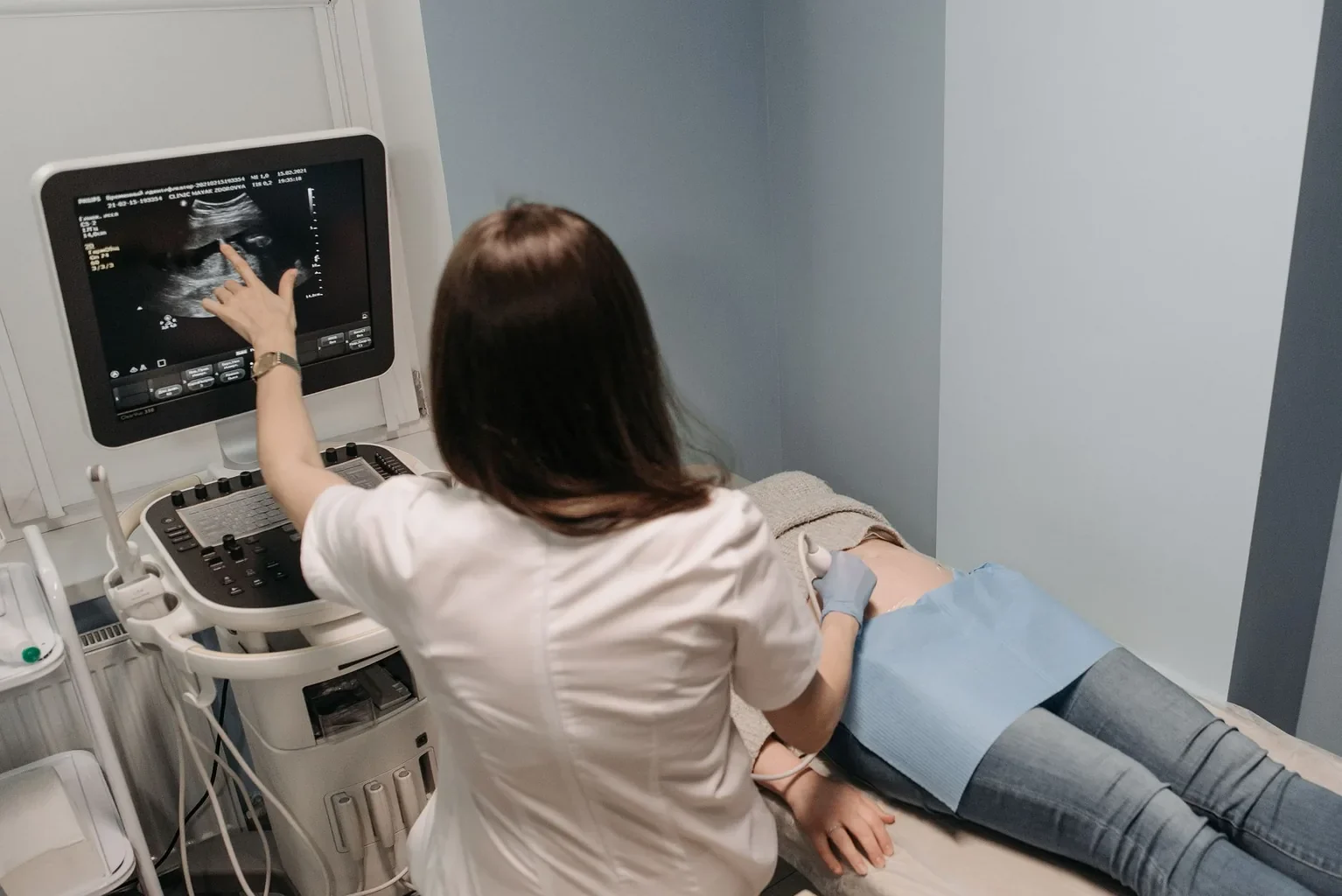
Abdominal ultrasound. The first-line imaging test in US clinical practice. Ultrasound is excellent at detecting gallstones, gallbladder wall thickening, and surrounding fluid (signs of inflammation). It is non-invasive, widely available, fast, and does not use radiation. According to NIDDK and major US guidelines, transabdominal ultrasound is the standard initial test.
HIDA scan (cholescintigraphy). A nuclear medicine test that evaluates gallbladder function. A small amount of radioactive tracer is injected; the gallbladder’s ability to fill, contract, and release bile is measured. Used when ultrasound is inconclusive or to assess gallbladder dyskinesia (functional dysfunction without stones). The “gallbladder ejection fraction” calculated from the scan helps make the diagnosis.
CT scan of the abdomen. Useful for assessing complications such as gallbladder perforation, abscess, or pancreatitis. Less sensitive than ultrasound for the gallstones themselves but better for surrounding anatomy.
Magnetic resonance cholangiopancreatography (MRCP). Detailed MRI imaging of the bile ducts and pancreatic duct. Used when bile duct stones (choledocholithiasis) or anatomic variations are suspected, or to plan therapy.
Endoscopic retrograde cholangiopancreatography (ERCP). Both diagnostic and therapeutic. Used when bile duct stones need to be removed before or after cholecystectomy. Less commonly used purely for diagnosis since MRCP is non-invasive.
Endoscopic ultrasound (EUS). Combines endoscopy with ultrasound. Reserved for specific situations like suspected small bile duct stones or pancreatic involvement.
Differential Considerations
Not everyone with upper right abdominal pain has gallbladder disease. The diagnostic workup may identify or rule out other conditions:
- Peptic ulcer disease or gastritis
- Gastroesophageal reflux disease (GERD)
- Pancreatitis (which can also be triggered by gallstones)
- Hepatitis or other liver diseases
- Kidney stones or pyelonephritis on the right side
- Right-sided pneumonia with referred upper abdominal pain
- Irritable bowel syndrome with right-upper-quadrant symptoms
- Atypical cardiac pain (rarely presents this way but worth considering in older adults)
- Functional dyspepsia
Some patients have gallstones plus another contributing condition. The imaging and lab workup help determine which conditions are causing symptoms.
When to See a Doctor
Schedule an evaluation with your primary care doctor or a gastroenterologist if you:
- Have recurring upper right or upper middle abdominal pain after meals
- Experience pain lasting more than an hour, especially with nausea
- Have family history of gallstones plus new digestive symptoms
- Have rapid weight loss with new abdominal symptoms
Seek urgent care or go to the emergency room if you have:
- Severe sudden abdominal pain that does not improve
- High fever and chills with abdominal pain
- Persistent vomiting that prevents drinking fluids
- Yellowing of skin or eyes (jaundice)
- Dark urine or pale-colored stools with abdominal pain
How the Diagnosis Is Made
The diagnosis of gallstones or gallbladder disease typically follows this process:
- Clinical evaluation: History and physical exam suggest gallbladder disease
- Blood work: Identifies infection, bile duct obstruction, or pancreatitis
- First-line imaging: Abdominal ultrasound confirms or excludes gallstones
- Specialized imaging (if needed): HIDA scan for functional issues, MRCP for bile duct stones, CT for complications
- Subspecialty referral: Patients with symptomatic gallstones are typically referred to a general surgeon for evaluation; complications may need gastroenterology or interventional radiology
- Decision on treatment: Watchful waiting for asymptomatic stones; surgical removal of the gallbladder (cholecystectomy) for symptomatic disease or complications
What Happens After Diagnosis
Once gallbladder disease is confirmed, the next step depends on symptoms and risk factors:
- Asymptomatic gallstones: Usually no treatment, just monitoring
- Symptomatic gallstones: Referral to a general surgeon for cholecystectomy evaluation
- Acute cholecystitis: Often hospital admission; surgery within days
- Bile duct stones or pancreatitis: May need ERCP plus subsequent cholecystectomy
- Gallbladder dyskinesia with severe symptoms: Surgical consultation; outcomes more variable
The surgical evaluation and procedure are covered in our cholecystectomy candidate evaluation article and procedure and recovery article.
Continue Reading the Gallstones Cluster
- Gallstones: Overview, Types, and Symptoms
- Gallstones: Treatment, Prevention, and Management
- Gallstones: FAQs, Statistics, and Patient Stories
- Cholecystectomy: Candidate Evaluation and When to Consider Surgery
Sources
- National Institute of Diabetes and Digestive and Kidney Diseases (NIDDK). Gallstones diagnosis. https://www.niddk.nih.gov/health-information/digestive-diseases/gallstones
- American College of Surgeons (ACS). Gallbladder disease workup. https://www.facs.org/
- American Society for Gastrointestinal Endoscopy (ASGE). ERCP and biliary endoscopy. https://www.asge.org/
- American Gastroenterological Association (AGA). Clinical practice updates on gallstones. https://www.gastro.org/
- Mayo Clinic. Gallstones: diagnosis. https://www.mayoclinic.org/diseases-conditions/gallstones/diagnosis-treatment/drc-20354220
- Cleveland Clinic. Gallstones. https://my.clevelandclinic.org/health/diseases/7313-gallstones
- Johns Hopkins Medicine. Biliary diseases. https://www.hopkinsmedicine.org/health/conditions-and-diseases/gallbladder-disease
- NIH MedlinePlus. Gallstones. https://medlineplus.gov/gallstones.html
Medical Disclaimer
The information in this article is for general education and is not a substitute for professional medical advice, diagnosis, or treatment. Always consult a qualified medical professional with questions about gallbladder disease, symptoms, or diagnostic testing.