Medically reviewed by the Know Your Surgery Editorial Team. Last reviewed: May 2026.
Once brain tumor removal is decided, most patients and families want to know exactly what to expect. Modern neurosurgery in the United States follows established pathways, with details that vary by tumor location and approach. This article walks through preparation, the surgery step by step, anesthesia, and what recovery and rehabilitation typically look like over the days, weeks, and months that follow.
If you have not yet read about why surgery may be needed, our causes, diagnosis, and decision article covers how the decision is made.
Preparing for Brain Tumor Removal
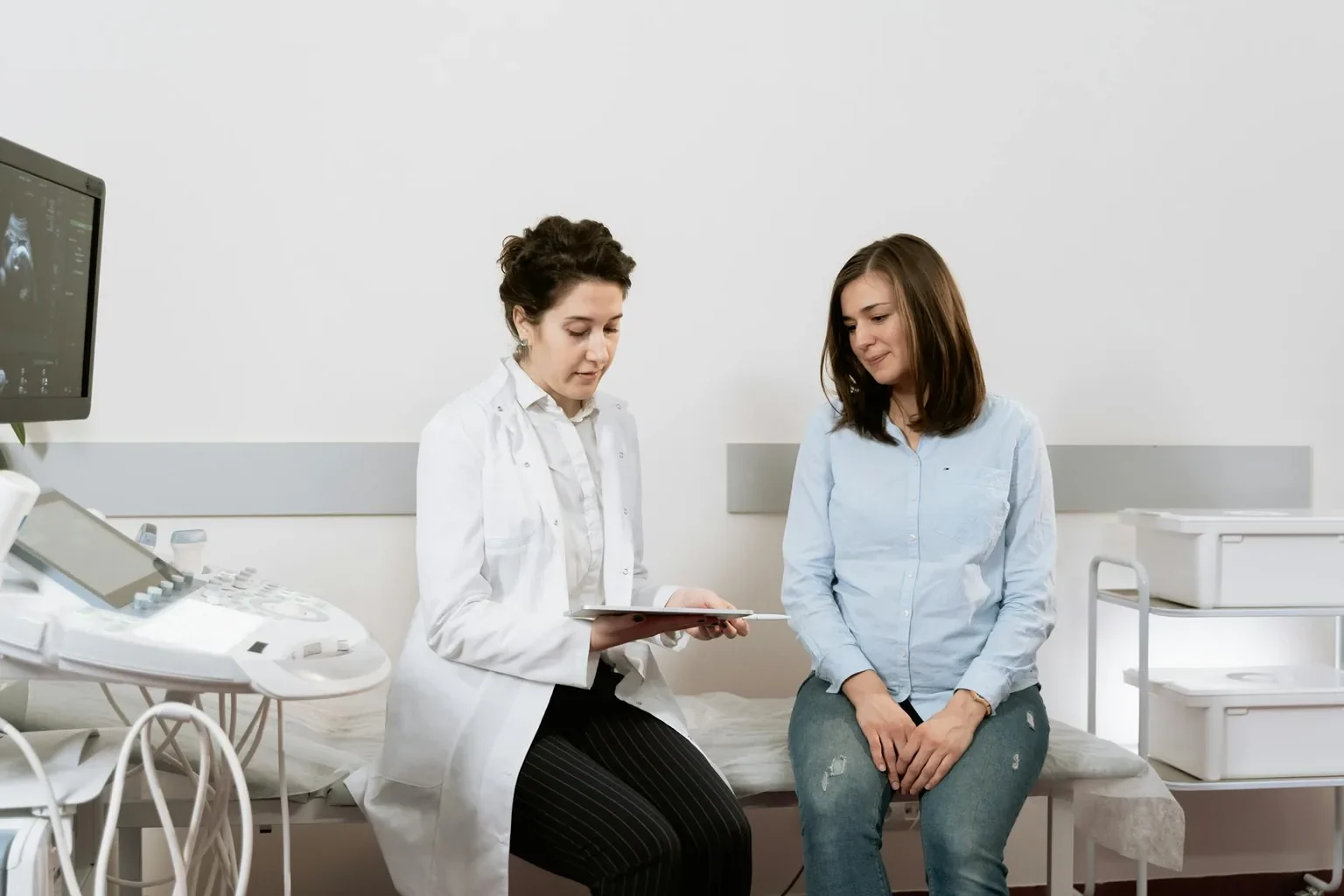
Brain tumor removal is usually scheduled several days to a few weeks in advance. The neurosurgical team typically:
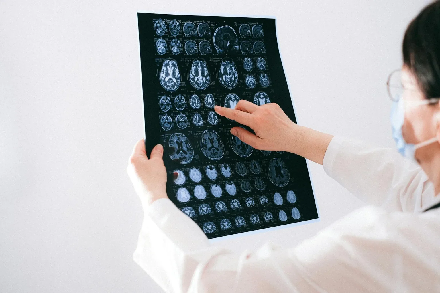
- Reviews recent imaging (MRI, fMRI, DTI as needed)
- Reviews medications, especially blood thinners
- Orders pre-operative blood tests
- Starts steroids (often dexamethasone) to reduce brain swelling
- Starts anti-seizure medication if needed
- Asks the patient to fast the night before surgery
- Performs an anesthesia consultation
- Discusses informed consent in detail
- Answers family questions about the day of surgery and the immediate recovery
The patient arrives at the hospital, changes into a gown, has IVs placed, and is brought to the operating room.
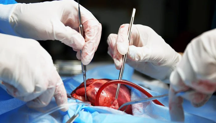
Craniotomy Step by Step
Craniotomy is the most common approach for tumors in the cerebrum or cerebellum. The full procedure typically takes 3 to 6 hours.
- General anesthesia is administered. The patient is fully asleep.
- The head is positioned and held still with a special head holder.
- A small section of hair is clipped at the surgical site, and the scalp is prepared with antiseptic.
- An incision is made in the scalp, and the scalp is gently moved aside.
- A small bone flap (a section of skull) is temporarily removed using a precise drill.
- The dura (covering of the brain) is opened carefully.
- Image guidance and intraoperative monitoring help locate the tumor and avoid critical brain structures.
- The tumor is removed in pieces using gentle suction, ultrasonic aspirators, or specialized instruments. The neurosurgeon takes care to preserve healthy brain tissue.
- The dura is closed, and the bone flap is replaced and secured with small titanium plates and screws.
- The scalp is closed in layers with sutures or staples.
- The patient is awakened and moved to a neurosurgical intensive care unit for close monitoring.
Awake Craniotomy Step by Step
Awake craniotomy is used for tumors near critical brain regions controlling speech or movement. The full procedure typically takes 4 to 6 hours.
- Sedation is given for the initial scalp incision and bone removal.
- The patient is awakened for the part of surgery near critical brain regions.
- A neuropsychologist or speech therapist asks the patient to speak, name objects, or move while the surgeon stimulates the brain to map function.
- The tumor is removed, with continuous testing to preserve speech and movement.
- Sedation resumes for closure.
The patient feels no pain because the brain itself has no pain receptors. The scalp is fully numbed.
Transsphenoidal Surgery Step by Step
Transsphenoidal surgery is used for pituitary tumors and some skull base tumors. The full procedure typically takes 2 to 4 hours.
- General anesthesia is administered.
- A thin endoscope is passed through one nostril to the back of the nose.
- The thin bone behind the nose is gently opened to reach the pituitary region.
- The tumor is removed with specialized instruments.
- Any opening in the dura is sealed with tissue or grafting material.
- No external incisions are needed.
Recovery is typically quicker than with craniotomy.
Stereotactic Biopsy Step by Step
Used for diagnosis when full removal is not the goal. Typically takes 1 to 2 hours.
- Local anesthesia or general anesthesia is used.
- A small frame or imaging system is used to guide a thin needle precisely.
- A small skin incision and tiny opening in the skull are made.
- A needle takes small tissue samples for pathology.
- The skin is closed with one or two stitches.
Most patients can go home the same day or the next morning.
Anesthesia for Brain Tumor Surgery
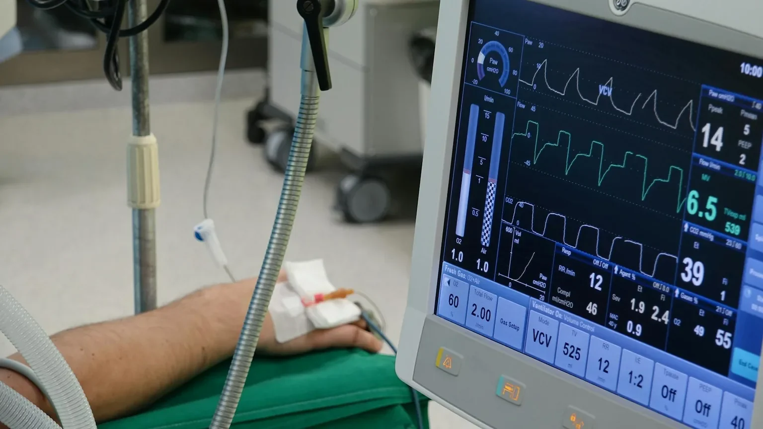
Most brain tumor surgeries use general anesthesia. Awake craniotomy uses a combination of sedation and a wakeful period. The anesthesia team monitors closely throughout, including breathing, heart rhythm, oxygen, and brain pressure.
Anesthesia for neurosurgery is highly specialized. The team accounts for any heart, lung, kidney, or other medical conditions in the plan.
What Happens Right After Surgery
After surgery, the patient is moved to a neurosurgical intensive care unit (NICU) for close monitoring of vital signs, pain, and neurological status (mental clarity, strength, vision, speech, movement). The team checks neurological status often.
Common features of the early hours:
- Headache and scalp soreness, controlled with medications
- Some swelling around the eyes, especially after surgeries near the front of the head
- Brief tiredness or confusion as anesthesia wears off
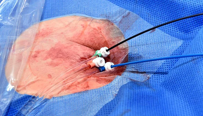
- A drain or stitches at the surgical site
- Compression devices on the legs to reduce blood-clot risk
A CT or MRI is typically done within a day to confirm the surgery went as planned and to check for bleeding or swelling.
Recovery Timeline
Recovery depends on the surgical approach, tumor type, and the patient’s pre-operative function.
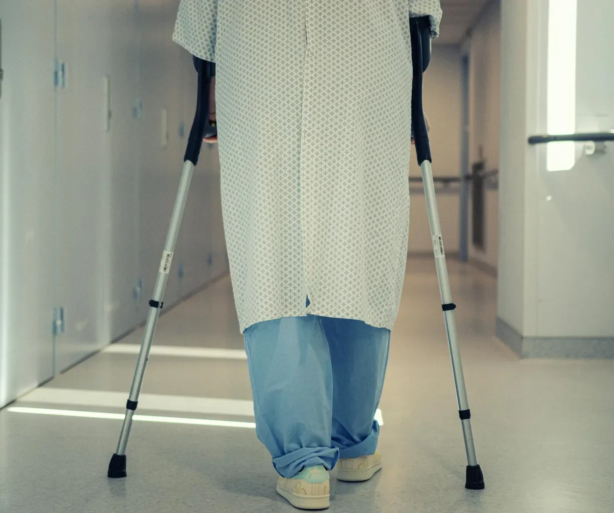
First 48 hours. ICU stay, neurological checks every 1 to 2 hours, pain control. Walking begins when safe (often the next morning) to reduce blood-clot risk.
Days 2 to 5. Move to a regular hospital room. Diet advances. Walking and basic activities of daily living resume. Steroids and anti-seizure medications are adjusted. The patient and family meet with the rehabilitation team if needed.
Days 3 to 7. Many patients are discharged home. Patients with rehabilitation needs may transfer to inpatient or outpatient therapy.
Weeks 1 to 4 at home. Rest, gradual return to light activity. No driving until cleared. No heavy lifting. Wound care and stitch or staple removal at the follow-up visit.
Weeks 4 to 12. Most patients return to office work and many normal activities, depending on the surgery. Outpatient rehabilitation continues if needed.
Months 3 to 12. Continued recovery, especially for patients with rehabilitation needs. Some symptoms continue to improve gradually for many months.
These timelines are general. Patients with complex surgery or rehabilitation needs may take longer. Always follow your neurosurgical team’s specific instructions.
Diet, Medications, and Wound Care
Most patients can return to a normal diet quickly after surgery. Specific instructions:
- Steroids are tapered over days to weeks based on swelling.
- Anti-seizure medications may be continued depending on history and surgery.
- Pain medications are used as needed; many patients transition to acetaminophen within days.
- Wound care focuses on keeping the incision clean and dry. Showering is usually allowed at the follow-up visit.
Pain Management
Pain after brain tumor removal is typically well controlled with:
- Scheduled doses of acetaminophen
- Short-term use of stronger pain medication for the first few days
- Cold packs to reduce swelling
- Adjustment of medications as needed
Severe headache, fever, drainage, or worsening neurological symptoms should prompt immediate contact with the surgical team.
Activity Restrictions and Common-Sense Care
In the first 1 to 6 weeks, the brain and skull are healing. Common restrictions include:
- No heavy lifting (typically over 10 to 15 pounds) for several weeks
- No driving until cleared by the surgical team and free of seizures
- No strenuous exercise until cleared
- No alcohol while on certain medications
- Walking is encouraged from early days
- No swimming or hot tubs until incisions are fully healed
Detailed warning signs are covered in our FAQs and statistics article.
Rehabilitation After Brain Tumor Removal
Many patients benefit from formal rehabilitation, especially if the tumor was near critical brain regions. Rehabilitation may include:
- Physical therapy for strength, balance, and walking
- Occupational therapy for daily activities and fine motor skills
- Speech therapy for speech, language, swallowing, and cognitive skills
- Neuropsychology for memory, attention, and emotional support
- Vocational rehabilitation for return to work
Rehabilitation can take place inpatient (for patients who need intensive support), at a skilled nursing or rehabilitation facility, or in outpatient settings.
Follow-Up Appointments
A typical follow-up schedule includes:
- Phone or in-person check during the first week
- Office visit at 2 weeks for stitch or staple removal and wound check
- Pathology review meeting to discuss the tumor type and any further treatment
- MRI scans at intervals (often 2 to 3 months at first, then less often)
- Neuro-oncology and radiation oncology visits if additional treatment is planned
For patients receiving radiation or chemotherapy, the follow-up calendar expands to include those visits.
Long-Term Outlook
Outcomes vary widely based on tumor type, grade, location, and patient health. Some patients (with low-grade tumors fully removed) live many years with minimal long-term effects. Others (with high-grade malignant tumors) need ongoing treatment and may face significant challenges. The neuro-oncology team guides expectations honestly and supports the patient and family throughout.
Continue Reading the Brain Tumor Removal Cluster
- Brain Tumor Removal: Overview, Types, and What to Expect
- Brain Tumor Removal: Causes, Diagnosis, and When to Consider Surgery
- Brain Tumor Removal: FAQs, Statistics, and Patient Stories
Sources
- American Association of Neurological Surgeons (AANS). Brain tumors: treatment. https://www.aans.org/patients/conditions/brain-tumors/
- National Cancer Institute (NCI). Adult central nervous system tumors: treatment. https://www.cancer.gov/types/brain
- Mayo Clinic. Brain tumor: treatment and recovery. https://www.mayoclinic.org/diseases-conditions/brain-tumor/diagnosis-treatment/drc-20350088
- Cleveland Clinic. Brain surgery: procedure and recovery. https://my.clevelandclinic.org/health/treatments/4078-brain-surgery
- American Society for Radiation Oncology (ASTRO). Patient resources. https://www.astro.org/patient-care-and-research/patient-education/
Medical Disclaimer
The information in this article is for general education and is not a substitute for professional medical advice, diagnosis, or treatment. Always consult a qualified healthcare provider with questions about your surgery, medications, or rehabilitation. Never disregard professional medical advice or delay in seeking it because of something you have read here. If you experience severe headache, fever, worsening neurological symptoms, seizure, or signs of infection after surgery, contact the surgical team immediately or go to the nearest emergency department.