Medically reviewed by the Know Your Surgery Editorial Team. Last reviewed: May 2026.
Kidney stone removal is recommended when a stone is unlikely to pass safely on its own, when it causes severe symptoms, or when it threatens kidney function. This article explains what causes kidney stones, how urologists confirm the diagnosis, and how the decision to proceed with kidney stone removal is made. If you are new to the topic, the cluster overview article is a useful starting place.
What Causes Kidney Stones?
Kidney stones form when the urine becomes too concentrated, allowing minerals to crystallize and clump together. Several specific causes are recognized in the United States.
Low fluid intake. Not drinking enough water is the single most common preventable cause. Concentrated urine allows minerals to crystallize.
Diet. Diets high in salt, animal protein, or oxalate-rich foods (such as spinach, nuts, and certain teas) can raise the risk in some patients. Sugary drinks, especially those with high-fructose corn syrup, are also associated with higher risk.
Genetics and family history. Some patients are born with metabolic differences (such as cystinuria or distal renal tubular acidosis) that raise their stone risk. Family history of stones is itself a risk factor.
Urinary tract conditions. Recurrent urinary tract infections, an obstructed urinary tract, or anatomical variations can raise stone risk.
Medical conditions. Obesity, diabetes, gout, hyperparathyroidism, inflammatory bowel disease, and gastric bypass surgery all raise stone risk.
Medications. Some medications (including certain diuretics, antiseizure medications, and HIV protease inhibitors) can contribute to stone formation.
Climate and dehydration. Living in hot, dry climates and sweating heavily without replacing fluids raises risk.
There are several stone types, each with somewhat different causes:
- Calcium oxalate is the most common type in the United States.
- Calcium phosphate is less common and may be linked to metabolic conditions.
- Uric acid stones are linked to gout, high-protein diets, and persistently acidic urine.
- Struvite stones form in patients with chronic urinary infections caused by certain bacteria.
- Cystine stones are rare and result from an inherited disorder.
Knowing the stone type, when possible, helps the team prevent future stones.
Risk Factors That Increase the Likelihood of Stones
Several factors are linked to a higher likelihood of stone disease:
- Age. Stones are most common between 30 and 60, although they can occur at any age.
- Sex. Slightly more common in men, but rates in women have been rising.
- Family history. Doubles the risk in many studies.
- Climate. Higher rates in the southeastern US (“the stone belt”).
- Body weight. Obesity raises risk.
- Past stones. A first stone strongly predicts future stones.
Knowing your risk factors helps you and your clinician choose preventive steps.
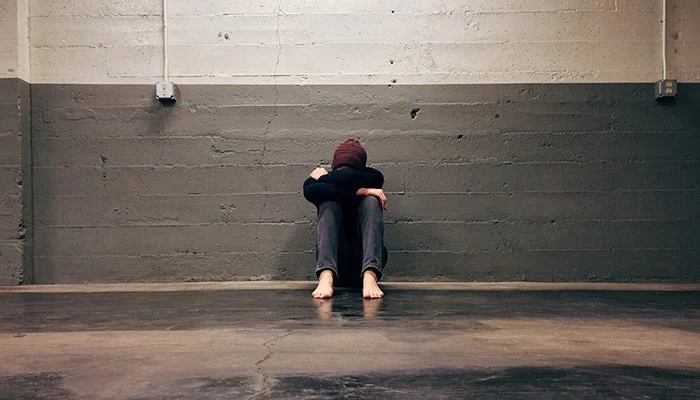
Symptoms Suggesting a Kidney Stone
Symptoms vary by stone size and location. Many small stones cause no symptoms at all and pass unnoticed. Larger or moving stones often produce a recognizable pattern.
Classic pattern:
- Sudden, severe pain in the back, side, or lower abdomen (“renal colic”)
- Pain that comes in waves and may radiate to the groin or testicles
- Restlessness (patients often pace because they cannot find a comfortable position)
- Pink, red, or brown urine (blood)
- Cloudy or foul-smelling urine
- Frequent, urgent urination
- Burning during urination
- Nausea and vomiting
Severe symptoms that require immediate emergency evaluation:
- High fever or shaking chills (possible infection above a blocked stone)
- Inability to urinate
- Severe vomiting that prevents fluid intake
- Fainting or signs of severe dehydration
Anyone with severe flank pain plus fever should seek immediate emergency evaluation. A blocked, infected stone (called obstructive pyelonephritis) is a urologic emergency that requires urgent drainage and antibiotics.
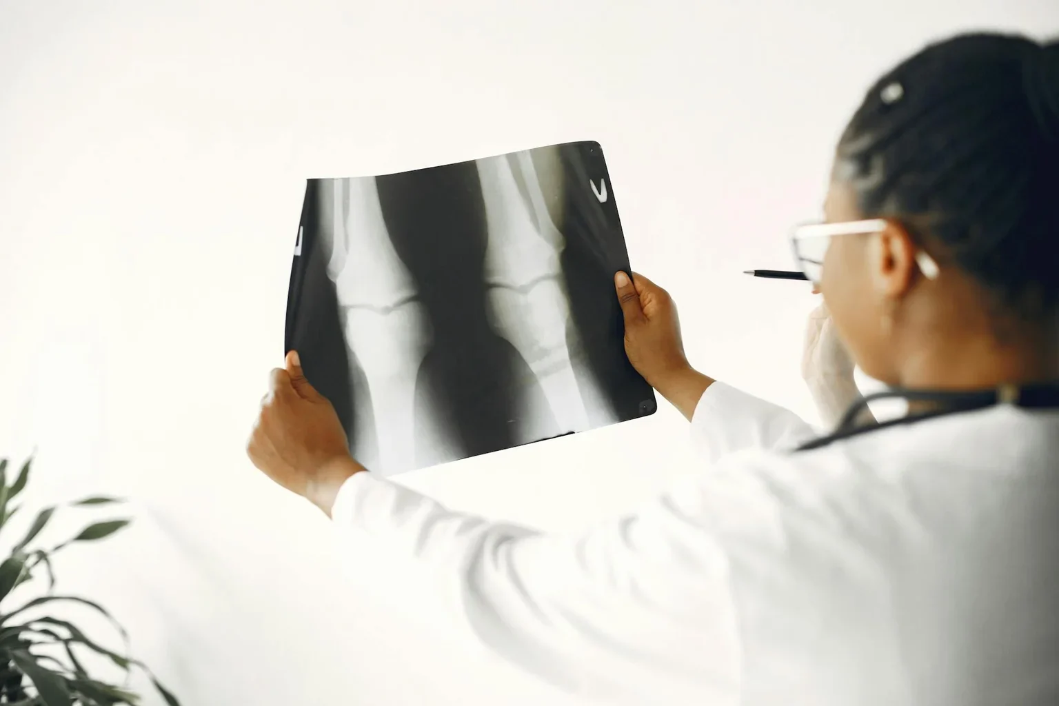
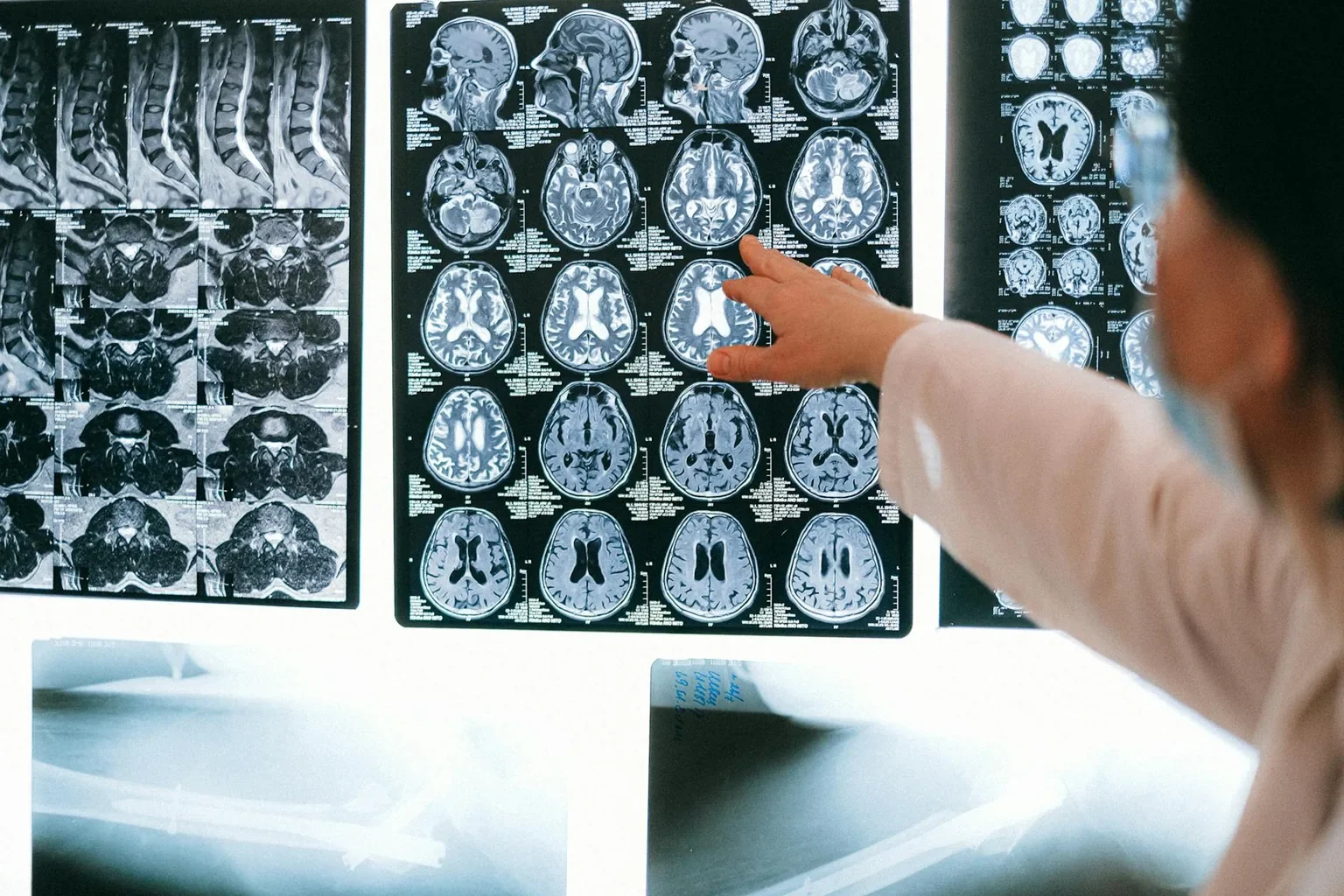
How Clinicians Diagnose Kidney Stones
When a patient arrives with possible stone symptoms, the team typically follows a structured evaluation.
Detailed history. The clinician asks about the timing, location, and character of the pain, prior stones, family history, fluid intake, diet, medications, and other medical conditions.
Physical examination. The clinician checks for tenderness in the back over the kidneys (called “costovertebral angle tenderness”) and abdominal tenderness, and looks for signs of infection (fever, fast heart rate).
Urinalysis. Looks for blood (almost always present with a stone), signs of infection, urine pH, and crystals.
Blood tests. A complete blood count and basic chemistry panel check for infection, kidney function, and metabolic disorders.
Imaging studies. The choice depends on the patient.
- Low-dose CT scan of the abdomen and pelvis is the most accurate test for stones in adults and is widely used in US emergency departments.
- Ultrasound is preferred in pregnant patients and often used as a first test in children to avoid radiation.
- KUB X-ray (kidneys-ureter-bladder) is sometimes used for follow-up of stones that show up on X-ray.
Stone analysis. If a stone is captured (passed naturally or removed during a procedure), it is sent to a lab to identify its mineral composition. This guides prevention.
24-hour urine collection. For patients with recurrent stones, a 24-hour collection measures urine volume, pH, and the levels of minerals that contribute to stone formation. This helps the team tailor preventive treatment.
Differential Diagnosis
Several conditions can mimic stone pain. The evaluation helps rule them out.
- Urinary tract infection or pyelonephritis
- Appendicitis (especially right-sided pain)
- Diverticulitis
- Ovarian cyst, ectopic pregnancy, or endometriosis
- Musculoskeletal back pain
- Aortic aneurysm (in older patients)
- Bowel obstruction
The diagnostic workup helps the team distinguish a stone from these conditions and choose the right next step.
Initial Management of Acute Stone Pain

Once a stone is confirmed, initial management focuses on:
- IV fluids for hydration
- Pain control (often with NSAIDs such as ketorolac, sometimes opioids)
- Anti-nausea medication
- Antibiotics if infection is present
- Medical expulsive therapy (a medication called tamsulosin that relaxes the ureter to help small stones pass)
- Stone strainer to catch the stone for analysis
Most small stones pass within a few days to a few weeks with this approach. Patients are typically asked to return to clinic for a follow-up scan.
When to Consider Kidney Stone Removal
Kidney stone removal is generally recommended when:
- The stone is too large to pass on its own (typically over 6 to 8 mm in adults)
- The stone is blocking urine flow from the kidney
- The stone causes severe pain that does not respond to medication
- The stone has not moved after weeks despite reasonable attempts at passage
- There is infection in the urinary tract along with a stone (urgent procedure)
- The patient has only one functioning kidney
- Stone is causing or worsening kidney damage
- The patient has a job (such as a pilot) where stone passage at the wrong moment could be unsafe
For some patients, the decision is shared between the urologist and the patient based on quality of life: how much pain the stone is causing, how disruptive it is, and how much time the patient is willing to wait for natural passage.
How the Decision Is Made
The choice of procedure (URS, SWL, or PCNL) depends on several factors:
Stone size and location. Small to medium stones in the ureter are usually treated with URS. Smaller stones in the kidney may respond well to SWL. Large stones (over 2 cm) and staghorn stones generally require PCNL.
Stone composition. Some stones (especially calcium oxalate monohydrate and cystine) are very hard and may not respond well to SWL. Imaging characteristics give the urologist clues about composition.
Patient anatomy and health. Body habitus, kidney shape, and prior surgeries can affect which approach is safe and effective.
Surgical urgency. A stone with infection is treated with urgent drainage (often a stent or nephrostomy tube), with definitive stone removal scheduled later.
Patient preference. Where multiple options are reasonable, the urologist discusses the pros and cons (recovery time, success rate, anesthesia type) with the patient.
Pre-Procedural Preparation
When kidney stone removal is planned, the team typically prepares the patient:
- Pre-operative blood and urine tests
- Medication review, including blood thinners and diabetes medications
- Anesthesia consultation
- Imaging review (CT or X-ray) on the day of the procedure
- Fasting as instructed (typically nothing to eat or drink after midnight)
- Antibiotics if a urinary infection is suspected
- Consent discussion about the procedure, alternatives, risks, and recovery
The procedure itself, the recovery, and the home recovery period are covered in detail in our procedure and recovery article.
Conditions That May Affect the Procedural Plan
Some coexisting conditions can change how kidney stone removal is performed:
- Pregnancy. SWL is avoided; ureteroscopy is generally preferred for stones requiring intervention.
- Bleeding disorders or anticoagulant therapy. Blood thinners may need to be paused; SWL is generally avoided.
- Severe obesity. May limit SWL effectiveness and may favor URS or PCNL.
- Single kidney. A more cautious approach may be chosen.
- Active urinary infection. Drainage with a stent or tube comes first; stone removal is delayed.
- Cardiac devices (pacemakers, defibrillators). May affect SWL planning.
The urology team accounts for these factors when choosing the safest approach.
What Happens After You Decide
Once kidney stone removal is scheduled, the urology team prepares the patient. The procedure itself, the recovery, and the home recovery period are covered in detail in our procedure and recovery article.
The conversation with your urologist is brief but important. Key things to confirm: the procedure plan (URS, SWL, or PCNL), whether a stent will be placed, expected recovery time, plan for stone analysis, and an emergency contact for after the procedure.
Continue Reading the Kidney Stone Removal Cluster
- Kidney Stone Removal: Overview, Types, and What to Expect
- Kidney Stone Removal: Procedure, Recovery, and Rehabilitation
- Kidney Stone Removal: FAQs, Statistics, and Patient Stories
Sources
- American Urological Association (AUA). Surgical management of stones guideline. https://www.auanet.org/guidelines-and-quality/guidelines
- National Institute of Diabetes and Digestive and Kidney Diseases (NIDDK). Kidney stones: causes and diagnosis. https://www.niddk.nih.gov/health-information/urologic-diseases/kidney-stones
- Centers for Disease Control and Prevention (CDC). Kidney disease statistics. https://www.cdc.gov/kidney-disease/data-research/
- Mayo Clinic. Kidney stones: diagnosis and treatment. https://www.mayoclinic.org/diseases-conditions/kidney-stones/diagnosis-treatment/drc-20355759
- Cleveland Clinic. Kidney stones: causes and diagnosis. https://my.clevelandclinic.org/health/diseases/15604-kidney-stones
Medical Disclaimer
The information in this article is for general education and is not a substitute for professional medical advice, diagnosis, or treatment. Always consult a qualified healthcare provider with questions about kidney pain or urinary symptoms. Never disregard professional medical advice or delay in seeking it because of something you have read here. If you have severe flank pain with fever, chills, or inability to urinate, seek immediate medical evaluation in an emergency department.